Class 12 : Biology (English) – Lesson 2: Human Reproduction
EXPLANATION & SUMMARY
🌱 Introduction
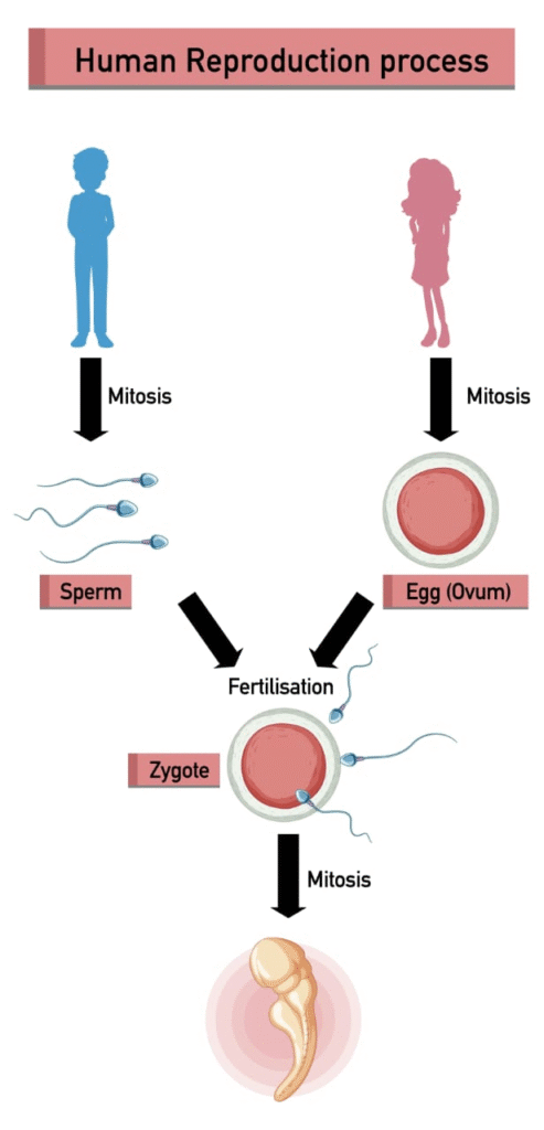
Reproduction is one of the fundamental characteristics of living organisms, ensuring the continuity of species. In humans, it is sexual, involving two parents, and it results in the formation of offspring through the process of gamete fusion (fertilisation), development of the embryo, and birth.
The process of human reproduction can be divided into the following stages: ➤ Gametogenesis
➤ Insemination
➤ Fertilisation
➤ Implantation
➤ Gestation
➤ Parturition
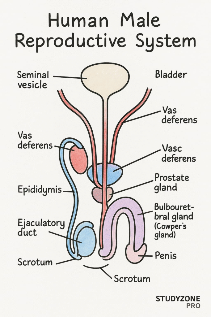
👨 Male Reproductive System
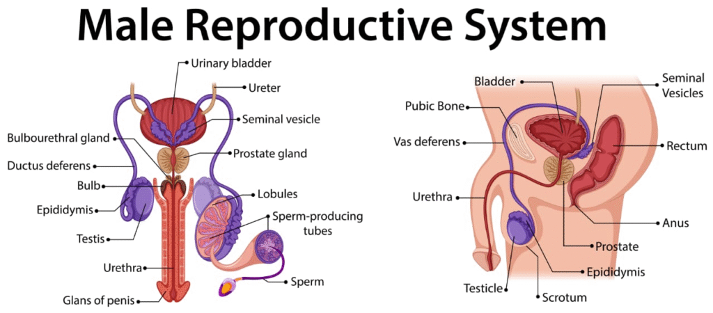
The male reproductive system consists of:
Pair of testes
Reproductive ducts
Accessory glands
External genitalia
Testes:
Each testis is oval-shaped, enclosed in a scrotum which keeps the testes 2–2.5°C below body temperature (necessary for spermatogenesis). Each testis has about 250 compartments called testicular lobules, and each lobule contains 1–3 seminiferous tubules, the site of sperm formation.
Seminiferous Tubules:
These have two types of cells:
Spermatogenic cells – develop into sperm
Sertoli cells – nourish the developing sperms
In between the tubules lie Leydig cells, which secrete androgens (mainly testosterone).
Duct System:
Sperm from seminiferous tubules are transported through: • Rete testis → Vasa efferentia → Epididymis → Vas deferens
Vas deferens unites with the duct of the seminal vesicle to form the ejaculatory duct, which opens into the urethra that runs through the penis.
Accessory Glands:
Seminal vesicles (secrete fructose-rich alkaline fluid)
Prostate gland (adds enzymes and calcium)
Bulbourethral glands (secrete mucus for lubrication)
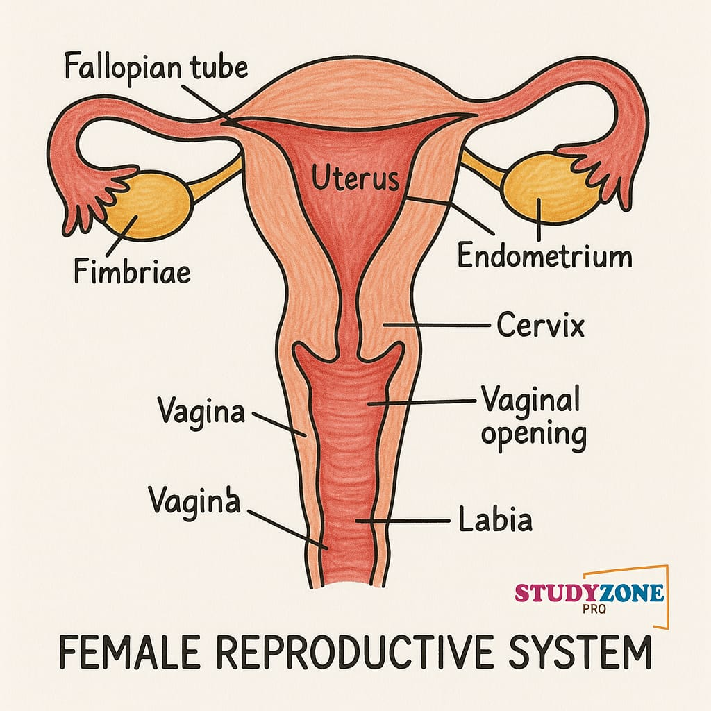
👩 Female Reproductive System
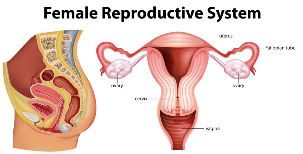
The female reproductive system consists of:
A pair of ovaries
A pair of oviducts (fallopian tubes)
A uterus
A vagina
External genitalia
Ovaries:
Primary female sex organs producing ova and hormones (estrogen, progesterone). Covered by germinal epithelium and divided into cortex and medulla.
Oviducts (Fallopian Tubes):
Each tube is 10–12 cm long and divided into:
Infundibulum with fimbriae to collect ova
Ampulla – site of fertilisation
Isthmus – narrow segment connecting to uterus
Uterus (Womb):
Pear-shaped, thick-walled organ lined with endometrium, muscular myometrium, and outer perimetrium.
Vagina:
Elastic muscular canal that receives the penis during intercourse and serves as the birth canal.
External Genitalia (Vulva):
Includes mons pubis, labia majora, labia minora, clitoris, and vaginal orifice.
Mammary Glands:
Modified sweat glands involved in lactation. Composed of lobes, lobules, alveoli (milk-secreting), ducts, and nipple.
🧬 Gametogenesis
🧪 Spermatogenesis:
Occurs in seminiferous tubules
Begins at puberty under hormonal control
Steps:
➤ Spermatogonia (2n) → primary spermatocytes
➤ Primary spermatocyte (2n) → meiosis I → 2 secondary spermatocytes (n)
➤ Meiosis II → 4 spermatids
➤ Spermiogenesis: Spermatids → Sperm
➤ Spermiation: Sperm released into tubule lumen
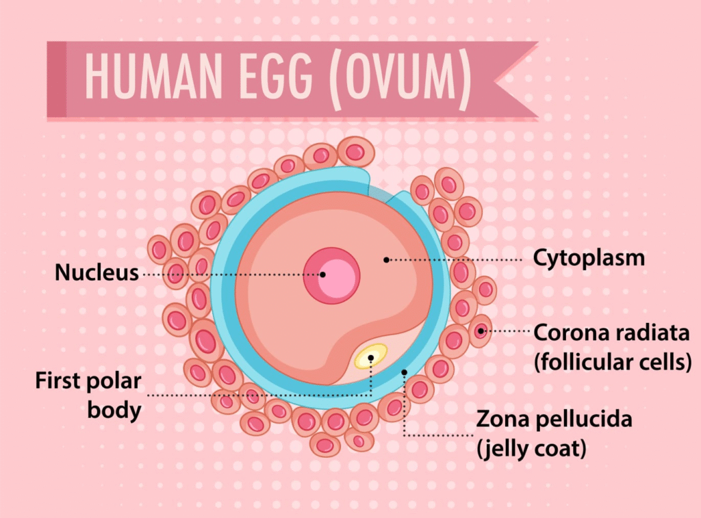
🧪 Oogenesis:
Begins during fetal life
Oogonia (2n) → primary oocytes (arrested in prophase I)
At puberty, one primary oocyte completes meiosis I each cycle
➤ Primary oocyte → secondary oocyte (n) + first polar body
➤ Secondary oocyte is ovulated and arrested in metaphase II
➤ Meiosis II completes only upon fertilisation → ovum + second polar body
💉 Hormonal Control of Gametogenesis
GnRH from hypothalamus → stimulates pituitary
Pituitary secretes:
➤ FSH – acts on Sertoli cells and ovarian follicles
➤ LH – triggers testosterone (in males) and ovulation (in females)
Inhibin (from Sertoli cells) → inhibits FSH
Estrogen & Progesterone → regulate female cycle and pregnancy
🧪 Menstrual Cycle
Cycle of ~28 days in human females.
Phases:
Menstrual Phase (Days 1–5):
Shedding of endometrium if fertilisation does not occur
Follicular Phase (Days 6–13):
FSH stimulates follicle growth; estrogen rebuilds endometrium
Ovulation (Day 14):
LH surge causes ovulation (release of secondary oocyte)
Luteal Phase (Days 15–28):
Corpus luteum secretes progesterone → maintains endometrium
If no fertilisation → corpus luteum degenerates → menstruation begins again
🧬 Fertilisation
Occurs in the ampullary-isthmic junction of fallopian tube.
Process:
Sperm travels through uterus and fallopian tube
Acrosome enzymes help sperm penetrate zona pellucida
Fusion of male and female gametes → zygote (2n)
Meiosis II completes in oocyte only after sperm entry
🌱 Implantation and Pregnancy
Zygote → undergoes cleavage → forms morula → blastocyst
Blastocyst has:
Trophoblast (outer layer) → forms placenta
Inner cell mass → develops into embryo
Implantation:
Blastocyst attaches to endometrium on 7th day post-fertilisation
🌾 Placenta and Embryonic Development
Placenta:
Temporary organ that connects embryo to uterine wall
Exchanges nutrients, gases, waste
Secretes hormones: hCG, hPL, estrogen, progesterone, relaxin
Embryonic Development: • Weeks 1–8: Embryo
After week 8: Fetus
All organs developed by 12 weeks
Movement by ~5 months
Lungs mature by 7th month
Fetus is fully developed by 9th month
🤱 Parturition (Childbirth)
Triggered by: ➤ Fetal hormones (like cortisol)
➤ Oxytocin – stimulates strong uterine contractions
➤ Relaxin – softens cervix
Process involves positive feedback loop: Uterine contraction → oxytocin → stronger contractions → delivery
🍼 Lactation
After parturition, prolactin stimulates milk production
Oxytocin aids in milk ejection or let-down reflex
Milk provides passive immunity to the infant via colostrum (rich in antibodies)
✍️ SUMMARY (~300 Words)
Humans reproduce sexually, involving the fusion of haploid gametes to form a diploid zygote.
The male reproductive system includes testes, ducts, glands, and penis. Spermatogenesis occurs in the seminiferous tubules. Sertoli cells provide nutrition; Leydig cells secrete testosterone.
The female reproductive system includes ovaries, fallopian tubes, uterus, vagina, and mammary glands. Oogenesis begins in fetal life and completes upon fertilisation.
The menstrual cycle (28 days) is regulated by FSH, LH, estrogen, and progesterone. Ovulation is induced by an LH surge.
Fertilisation occurs in the ampullary-isthmic junction. Sperm penetrates the ovum via acrosomal enzymes, forming a zygote.
The zygote undergoes cleavage to form a blastocyst, which implants into the uterus. This marks the beginning of pregnancy.
The placenta forms from trophoblast cells and enables nutrient/gas exchange. It secretes hormones like hCG, hPL, and progesterone.
The embryo develops into a fetus by the 9th week. Organs develop by 12 weeks, and full fetal development is complete by 9 months.
Parturition is initiated by hormonal signals including oxytocin and relaxin. It involves strong uterine contractions and results in childbirth.
Lactation is controlled by prolactin and oxytocin. Initial milk called colostrum provides immunity to the newborn.
This chapter lays the foundation for understanding reproductive physiology, hormonal control, and human developmental biology.
————————————————————————————————————————————————————————————————————————————
QUESTIONS FROM TEXTBOOK
Question 1. Fill in the blanks:
(a) Humans reproduce sexually
(b) Humans are viviparous
(c) Fertilisation is internal in humans
(d) Male and female gametes are haploid
(e) Zygote is diploid
(f) The process of release of ovum from a mature follicle is called ovulation
(g) Ovulation is induced by a hormone called LH (Luteinising Hormone)
(h) The fusion of male and female gametes is called fertilisation
(i) Fertilisation takes place in fallopian tube (oviduct)
(j) Zygote divides to form blastocyst which is implanted in uterus
(k) The structure which provides vascular connection between foetus and uterus is called placenta
Question 2. Draw a labelled diagram of male reproductive system.
Answer: Please refer to NCERT textbook diagram (Figure 2.1). A proper labelled diagram includes:
Testis
Epididymis
Vas deferens
Seminal vesicle
Prostate gland
Urethra
Penis
Scrotum
Question 3. Draw a labelled diagram of female reproductive system.
Answer: Please refer to NCERT textbook diagram (Figure 2.2). It should include:
Ovaries
Fallopian tubes
Uterus
Cervix
Vagina
Fimbriae
Endometrium
Question 4. Write two major functions each of testis and ovary.
Testis:
Produces male gametes (sperms) through spermatogenesis.
Secretes androgens (mainly testosterone) which regulate male secondary sexual characters and reproductive functions.
Ovary:
Produces female gametes (ova) through oogenesis.
Secretes female sex hormones (estrogen and progesterone), which regulate menstrual cycle and pregnancy.
Question 5. Describe the structure of a seminiferous tubule.
Answer:
Seminiferous tubules are coiled structures located in the testes, where sperm production occurs.
Structure:
Lined with a germinal epithelium that consists of two types of cells:
Spermatogenic cells – which form sperms in different stages of development.
Sertoli cells – which provide nourishment to developing sperms.
The space between tubules contains Leydig cells (interstitial cells) that produce androgens (testosterone).
Question 6. What is spermatogenesis? Briefly describe the process of spermatogenesis.
Answer:
Spermatogenesis is the process of formation of male gametes (sperms) in the seminiferous tubules of the testes. It occurs in the following stages:
Multiplication phase:
Spermatogonia (diploid, 2n) undergo mitosis to form more spermatogonia. Some grow into primary spermatocytes.
Growth phase:
Primary spermatocytes (2n) grow in size.
Maturation phase:
Primary spermatocytes undergo meiosis I to form two secondary spermatocytes (haploid, n).
Each secondary spermatocyte undergoes meiosis II to form two spermatids (haploid, n).
So, one primary spermatocyte gives rise to four spermatids.
Spermiogenesis:
Spermatids transform into mature spermatozoa (sperms).
Question 7. Name the hormones involved in regulation of spermatogenesis.
Answer:
The hormones regulating spermatogenesis are:
Gonadotropin-releasing hormone (GnRH): Secreted by the hypothalamus; it stimulates the anterior pituitary.
Follicle Stimulating Hormone (FSH): Stimulates Sertoli cells to support spermatogenesis.
Luteinising Hormone (LH): Stimulates Leydig cells to produce androgens (mainly testosterone).
Testosterone: Promotes sperm maturation and development of secondary sexual characteristics.
Inhibin: Secreted by Sertoli cells, it regulates FSH secretion via negative feedback.
Question 8. Define spermiogenesis and spermiation.Answer:
Spermiogenesis is the process of transformation of non-motile, round spermatids into mature, motile spermatozoa (sperms). This includes development of tail, condensation of nucleus, formation of acrosome, and removal of excess cytoplasm.
Spermiation is the process by which mature spermatozoa are released from the Sertoli cells into the lumen of the seminiferous tubule.
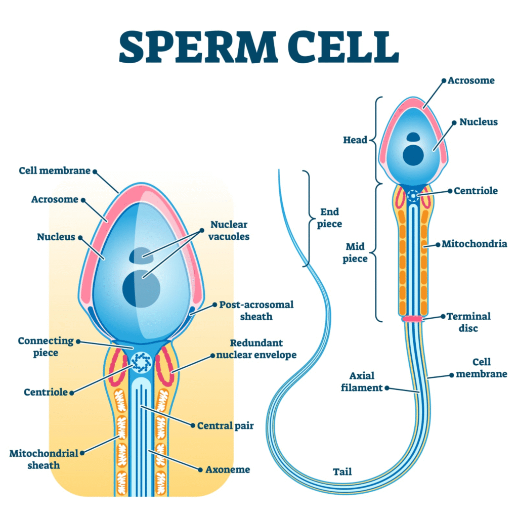
Question 9. Draw a labelled diagram of sperm.
Answer: Refer to NCERT Figure 2.3. Labelled parts include:
Head (nucleus + acrosome)
Neck
Middle piece (with mitochondria)
Tail
Question 10. What are the major components of seminal plasma?
Answer:
Seminal plasma is the fluid part of semen and is rich in:
Fructose – provides energy to sperms.
Calcium and enzymes – help in sperm motility and viability.
Prostaglandins – aid in sperm transport in female reproductive tract.
Buffers – maintain pH for sperm survival.
It is secreted mainly by:
Seminal vesicles (60%)
Prostate gland
Bulbourethral glands
Question 11. What are the major functions of male accessory ducts and glands?
Answer:
Accessory Ducts:
Epididymis: Storage and maturation of sperms.
Vas deferens: Transports sperms from epididymis to urethra.
Ejaculatory duct: Conveys sperms and secretions of glands into urethra.
Accessory Glands:
Seminal vesicles: Secrete fructose-rich alkaline fluid, the main part of semen.
Prostate gland: Adds enzymes and calcium, enhances sperm motility.
Bulbourethral glands: Secrete mucus that lubricates and neutralizes acidic urine traces in urethra.
Question 12. What is oogenesis? Give a brief account of oogenesis.
Answer:
Oogenesis is the process of formation of female gametes (ova) in the ovaries.
Stages:
Foetal Stage:
Oogonia (2n) divide mitotically to form millions of primary oocytes (2n), which begin meiosis I but get arrested in prophase I.
Puberty Onward:
Each menstrual cycle, a few primary oocytes resume meiosis. Only one completes meiosis I to form:
Secondary oocyte (n)
First polar body (n)
Secondary oocyte begins meiosis II but is arrested in metaphase II.
Fertilization:
Meiosis II completes only if sperm fertilizes the ovum, forming:
Ovum (n)
Second polar body (n)
Question 13. Draw a labelled diagram of a section through ovary.
Answer: Refer to NCERT Figure 2.4. Diagram should include:
Germinal epithelium
Cortex and medulla
Primary follicle
Secondary follicle
Tertiary follicle
Graafian follicle
Corpus luteum
(Use textbook or biology practical book for accurate drawing.)
Question 14. Draw a labelled diagram of a Graafian follicle.
Answer: Refer to NCERT Figure 2.5. Labels should include:
Secondary oocyte
Zona pellucida
Corona radiata
Antrum
Theca interna and externa
Cumulus oophorus
Follicular cells
(Kindly draw it neatly in your notebook as diagrams are not included here.)
Question 15. Name the functions of the following:(a) Corpus luteum:
Secretes progesterone which maintains the endometrium and supports implantation and early pregnancy.
(b) Endometrium:
Inner lining of the uterus; undergoes cyclic changes and is essential for implantation of the blastocyst.
(c) Acrosome:
Cap-like structure in the sperm head that contains hydrolytic enzymes (like hyaluronidase) to digest the egg coverings during fertilisation.
(d) Sperm tail:
Provides motility to the sperm, enabling it to swim toward the ovum.
(e) Fimbriae:
Finger-like projections at the end of fallopian tubes that help in capturing the ovum released from the ovary.
Question 16. Identify True/False statements. Correct each false statement to make it true.
(a) Androgens are produced by Sertoli cells. – False
Correction: Androgens are produced by Leydig cells, not Sertoli cells.
(b) Spermatozoa get nutrition from Sertoli cells. – True
(c) Leydig cells are found in ovary. – False
Correction: Leydig cells are found in testes, not ovary.
(d) Leydig cells synthesise androgens. – True
(e) Oogenesis takes place in corpus luteum. – False
Correction: Oogenesis takes place in the ovary, not corpus luteum.
(f) Menstrual cycle ceases during pregnancy. – True
(g) Presence or absence of hymen is not a reliable indicator of virginity or sexual experience. – True
Question 17. What is menstrual cycle? Which hormones regulate menstrual cycle?
Answer:
The menstrual cycle is a series of cyclic changes in the female reproductive system (mainly uterus and ovary) that prepares the body for pregnancy. It usually lasts for 28 days and includes:
Menstrual phase (Days 1–5): Shedding of endometrial lining.
Follicular phase (Days 6–13): Follicle growth and endometrium repair.
Ovulation (Day 14): Release of mature ovum from ovary.
Luteal phase (Days 15–28): Corpus luteum forms and secretes progesterone to maintain endometrium.
Hormones involved:
GnRH from hypothalamus
FSH and LH from anterior pituitary
Estrogen and progesterone from ovary
Question 18. What is parturition? Which hormones are involved in induction of parturition?
Answer:
Parturition is the process of childbirth or delivery of the baby from the uterus at the end of pregnancy.
Hormones involved:
Oxytocin: Secreted by maternal posterior pituitary; induces uterine contractions.
Relaxin: Helps in softening the cervix and relaxation of pelvic ligaments.
Cortisol (from fetus): Matures fetal organs and helps initiate labour.
Estrogens: Promote uterine sensitivity to oxytocin.
(Positive feedback mechanism between uterine contractions and oxytocin secretion.)
Question 19. In our society the women are often blamed for giving birth to daughters. Can you explain why this is not correct?
Answer:
This belief is scientifically incorrect and socially unjust. Sex of the child is determined by the type of sperm that fertilizes the ovum:
All ova carry an X chromosome.
Sperms can carry either X or Y chromosome.
If an X-bearing sperm fertilizes the ovum → female (XX)
If a Y-bearing sperm fertilizes the ovum → male (XY)
Thus, it is the male gamete (sperm) that determines the sex of the baby, not the female.
Question 20. How many eggs are released by a human ovary in a month? How many eggs do you think would have been released if the mother gave birth to identical twins? Would your answer change if the twins born were fraternal?
Answer:
Normally, one egg is released per month.
Identical twins arise from one fertilised egg (zygote) that splits into two embryos. So, only one ovum is released.
Fraternal twins are formed when two separate eggs are fertilised by two different sperms. So, in that case, two ova would have been released.
Question 21. How many eggs do you think were released by the ovary of a female dog which gave birth to 6 puppies?
Answer:
Dogs are polytocous animals (give birth to multiple offspring at once).
To give birth to 6 puppies, the female dog must have released 6 ova, each fertilised by a separate sperm.
Therefore, approximately 6 eggs were released.
.
————————————————————————————————————————————————————————————————————————————
OTHER IMPORTANT QUESTIONS FOR EXAMS
(CBSE MODEL QUESTIONS PAPER)
ESPECIALLY MADE FROM THIS LESSON ONLY
Q1. In human males, the urethra carries
(A) urine only
(B) semen only
(C) both urine and semen
(D) neither urine nor semen
Answer: (C) both urine and semen
Q2. Which of the following hormones triggers ovulation in human females?
(A) FSH
(B) LH
(C) Estrogen
(D) Progesterone
Answer: (B) LH
Q3. In which part of the female reproductive system does fertilisation normally occur?
(A) Vagina
(B) Uterus
(C) Fallopian Tube
(D) Ovary
Answer: (C) Fallopian Tube
Q4. The acrosome of a sperm is derived from which cell organelle?
(A) Nucleus
(B) Mitochondria
(C) Golgi body
(D) Ribosome
Answer: (C) Golgi body
Q5. Identify the correct pair:
(A) Sertoli cells – nutrition to sperm
(B) Leydig cells – produce FSH
(C) Graafian follicle – secretes testosterone
(D) Corpus luteum – produces FSH
Answer: (A) Sertoli cells – nutrition to sperm
Q6. Which layer of the uterus is shed during menstruation?
(A) Perimetrium
(B) Myometrium
(C) Endometrium
(D) Epimetrium
Answer: (C) Endometrium
Q7. Sperm maturation occurs in:
(A) Vas deferens
(B) Epididymis
(C) Ureter
(D) Urethra
Answer: (B) Epididymis
Q8. Menstrual flow occurs due to sudden reduction of which hormones?
(A) LH and FSH
(B) Estrogen and Progesterone
(C) Testosterone and LH
(D) LH and Progesterone
Answer: (B) Estrogen and Progesterone
Q9. Identify the function of the prostate gland:
(A) Produces sperm
(B) Provides nourishment to sperm
(C) Adds fluid for sperm motility
(D) Secretes testosterone
Answer: (C) Adds fluid for sperm motility
Q10. Ovulation typically occurs on which day of a 28-day menstrual cycle?
(A) Day 7
(B) Day 14
(C) Day 21
(D) Day 28
Answer: (B) Day 14
Q11. Number of chromosomes in a secondary oocyte:
(A) 46
(B) 23
(C) 92
(D) 44
Answer: (B) 23
Q12. Which is not a part of the human male reproductive system?
(A) Seminal vesicle
(B) Fallopian tube
(C) Vas deferens
(D) Epididymis
Answer: (B) Fallopian tube
Q13. The entry of sperm induces completion of:
(A) Mitosis
(B) First meiotic division
(C) Second meiotic division
(D) Cleavage
Answer: (C) Second meiotic division
Q14. Assertion (A): Corpus luteum secretes progesterone.
Reason (R): Progesterone helps in maintaining pregnancy.
(A) Both A and R are true; R explains A.
(B) Both A and R are true; R does not explain A.
(C) A is true, R is false.
(D) Both A and R are false.
Answer: (A) Both A and R are true; R explains A.
Q15. Assertion (A): Epididymis helps in the storage and maturation of sperm.
Reason (R): Epididymis secretes testosterone.
(A) Both A and R are true; R explains A.
(B) Both A and R are true; R does not explain A.
(C) A is true, R is false.
(D) Both A and R are false.
Answer: (C) A is true, R is false.
Q16. Identify the odd one out:
(A) Seminiferous tubules
(B) Vas deferens
(C) Prostate gland
(D) Fallopian tube
Answer: (D) Fallopian tube
Q17. Case-Based MCQ:
Read the following and answer:
During the menstrual cycle, the levels of LH peak sharply around mid-cycle, leading to the release of the ovum. The endometrial lining prepares for implantation.
What happens if fertilisation does not occur?
(A) Endometrium remains thick
(B) Progesterone levels rise further
(C) Endometrial lining breaks down
(D) LH remains high
Answer: (C) Endometrial lining breaks down
Q18. Case-Based MCQ:
During spermatogenesis, primary spermatocytes undergo meiosis to form haploid cells.
Which cells result directly from the first meiotic division?
(A) Spermatids
(B) Secondary spermatocytes
(C) Spermatozoa
(D) Spermatogonia
Answer: (B) Secondary spermatocytes
Q19. Write the role of oxytocin during parturition.
Answer:
Oxytocin stimulates strong uterine contractions during parturition (childbirth). These contractions help in the expulsion of the fetus from the uterus through the birth canal.
Q20. How does zona pellucida help in preventing polyspermy?
Answer:
After the first sperm successfully penetrates the zona pellucida, it undergoes chemical changes (zona reaction) which hardens the zona pellucida. This prevents the entry of additional sperms, thus avoiding polyspermy.
Q21. What is colostrum? Mention its significance.
Answer:
Colostrum is the first yellowish milk produced by the mother after childbirth. It is rich in antibodies (mainly IgA) and provides immunity to the newborn against infections.
Q22. How is the sex of a child determined in humans?
Answer:
The sex of a child is determined by the type of sperm that fertilizes the ovum.
Sperm carrying X chromosome → Female child (XX)
Sperm carrying Y chromosome → Male child (XY)
Since females produce only X-bearing eggs, the male determines the sex of the offspring.
Q23. List two major functions of placenta during pregnancy.
Answer:
It facilitates the exchange of nutrients, gases, and waste products between mother and fetus.
It acts as an endocrine organ by secreting hormones like hCG, estrogen, and progesterone to maintain pregnancy.
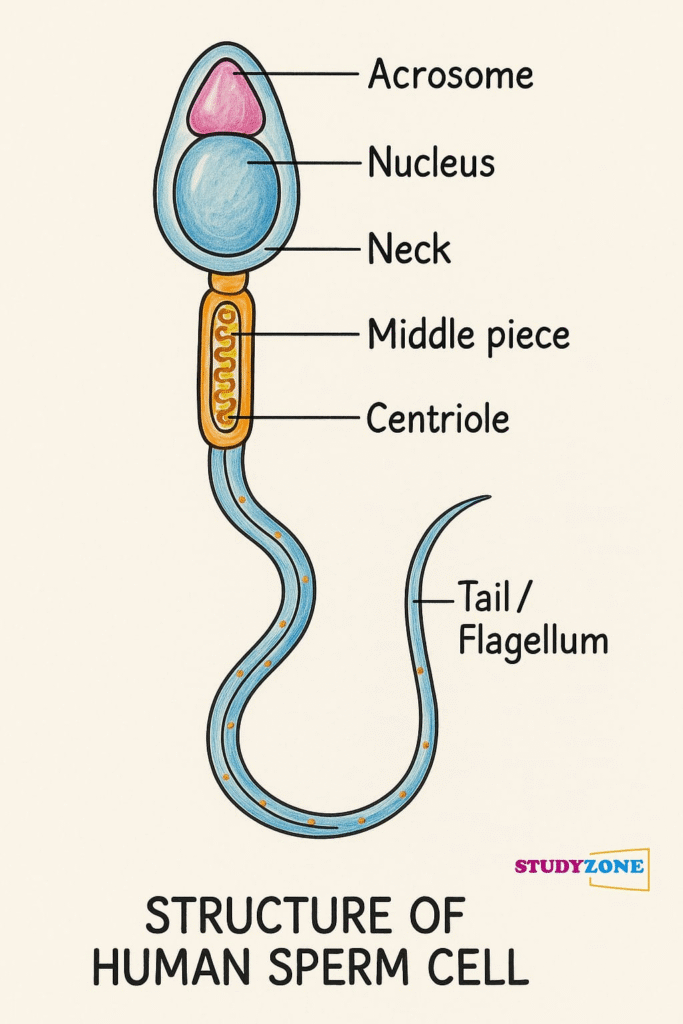
Q24. Describe the structure of a human sperm with the help of a labelled diagram.
Answer:
Structure of Human Sperm:
Head: Contains the nucleus with haploid chromosomes and is covered by the acrosome which contains enzymes for penetrating the egg.
Middle Piece: Contains mitochondria arranged spirally around the axial filament, providing energy for movement.
Tail: Long and slender, helps in the motility of the sperm.
(Labelled diagram should be drawn by student: Head → Middle Piece → Tail with clear parts marked)
Q25. Explain menstrual cycle phases briefly with key hormonal changes.
Answer:
Menstrual Phase (Days 1–5): Endometrium sheds; low levels of estrogen and progesterone.
Follicular Phase (Days 6–13): FSH stimulates follicle growth; estrogen levels rise, endometrium thickens.
Ovulation (Day 14): LH surge causes release of ovum from Graafian follicle.
Luteal Phase (Days 15–28): Corpus luteum forms, secretes progesterone to maintain endometrium.
Q26. Differentiate between spermatogenesis and oogenesis in three points.
Answer:
Feature Spermatogenesis Oogenesis
Site Seminiferous tubules (testes) Ovaries
Time of Initiation Puberty Fetal stage
Final Products 4 functional sperms 1 ovum and 2–3 polar bodies
Q27. Write the names and functions of any three hormones involved in the female reproductive cycle.
Answer:
FSH (Follicle Stimulating Hormone): Stimulates growth of ovarian follicles.
LH (Luteinizing Hormone): Triggers ovulation and formation of corpus luteum.
Progesterone: Prepares and maintains the endometrium for implantation.
Q28. Explain the process of fertilisation in humans.
Answer:
Sperm reaches the ampullary-isthmic junction of the fallopian tube where fertilisation occurs.
Acrosomal enzymes help sperm penetrate corona radiata and zona pellucida.
The sperm and ovum membranes fuse; sperm nucleus enters the ovum.
Meiosis II of ovum completes; male and female pronuclei fuse to form the diploid zygote.
Q29. Case-Based Question:
Read the following carefully and answer the questions below:
In humans, gamete formation is a complex process involving meiosis. Male gametes are produced in large numbers continuously after puberty, whereas female gametes are formed much earlier but complete their development later.
(i) Name the process of gamete formation in males and females.
(ii) Where does each of these processes occur?
(iii) Mention the ploidy of the gametes produced.
(iv) Give one difference between the timing of these processes in males and females.
Answer:
(i) Male: Spermatogenesis; Female: Oogenesis
(ii) Spermatogenesis occurs in seminiferous tubules of testes; Oogenesis occurs in ovaries.
(iii) Gametes produced are haploid (n).
(iv) Spermatogenesis starts at puberty and continues lifelong; Oogenesis starts during fetal development and pauses till puberty, completing only after fertilisation.
Q30. Case-Based Question:
Read the following carefully and answer the questions below:
The menstrual cycle is controlled by hormones from the pituitary and ovary. Disruptions in hormone levels can disturb the cycle.
(i) Which hormones from the pituitary are involved?
(ii) What are the roles of estrogen and progesterone?
(iii) What happens to the uterine lining if fertilisation does not occur?
(iv) Name the structure that secretes progesterone after ovulation.
Answer:
(i) FSH and LH from the pituitary.
(ii) Estrogen: Thickens endometrium; Progesterone: Maintains it for implantation.
(iii) Uterine lining breaks down, resulting in menstruation.
(iv) Corpus luteum.
Q31. Case-Based Question:
Read the passage and answer the following:
A newly fertilised egg travels through the fallopian tube to the uterus where it implants and starts forming an embryo. Several events occur during this period which ensure proper development.
(i) Name the process of attachment of the embryo to the uterine wall.
(ii) What forms the placenta?
(iii) State any two functions of the placenta.
(iv) Which hormone confirms pregnancy in a woman’s urine?
Answer:
(i) Implantation
(ii) Chorionic villi of the embryo and uterine tissue
(iii) Exchange of nutrients and gases; Secretes hormones like hCG
(iv) hCG (Human Chorionic Gonadotropin)
Q32. Explain the process of spermatogenesis in human males with a labelled diagram.
Answer:
Process of Spermatogenesis:
Spermatogonia (2n): Divide by mitosis, some differentiate into primary spermatocytes.
Primary Spermatocytes (2n): Undergo meiosis I to form two secondary spermatocytes (n).
Secondary Spermatocytes (n): Undergo meiosis II to form four spermatids (n).
Spermatids: Differentiate into spermatozoa through spermiogenesis.
Labelled Diagram:
(Seminiferous tubule cross-section showing stages: spermatogonia → primary spermatocyte → secondary spermatocyte → spermatid → spermatozoa.)
Final Products: 4 functional haploid sperm from each primary spermatocyte.
Q33. Describe the role of hormones during pregnancy and parturition.
Answer:
During Pregnancy:
hCG: Maintains corpus luteum, supports progesterone production.
Progesterone: Maintains endometrium, inhibits uterine contractions.
Estrogen: Supports uterine growth, breast development.
hPL (Human Placental Lactogen): Prepares mammary glands, regulates metabolism.
Relaxin: Softens cervix, loosens pelvic ligaments.
During Parturition:
Oxytocin: Stimulates strong uterine contractions for delivery.
Positive feedback loop: Contractions → more oxytocin → stronger contractions.
Progesterone declines; estrogen promotes uterine sensitivity to oxytocin.
Q34. Explain the structure and functions of the human female reproductive system with a neat, labelled diagram.
Answer:
Structure:
Ovaries: Produce ova and hormones (estrogen, progesterone).
Fallopian Tubes (Oviducts): Site of fertilisation; transport ovum.
Uterus: Thick muscular organ for implantation and fetal development.
Cervix: Connects uterus to vagina; controls passage.
Vagina: Birth canal, copulatory organ.
Functions:
Oogenesis, fertilisation, implantation, pregnancy support, parturition.
Diagram: Clearly labelled uterus, fallopian tubes, ovaries, cervix, vagina.
Q35. Describe the process of oogenesis with neat, labelled stages.
Answer:
Stages of Oogenesis:
Multiplication Phase (Fetal stage): Oogonia (2n) divide mitotically to form primary oocytes (2n).
Growth Phase: Primary oocytes grow in size.
Maturation Phase:
Primary oocyte completes meiosis I → secondary oocyte (n) + first polar body.
Secondary oocyte starts meiosis II, arrested at metaphase II until fertilisation.
On fertilisation, meiosis II completes → ovum (n) + second polar body.
Diagram: Clearly show oogonium → primary oocyte → secondary oocyte → ovum stages.
————————————————————————————————————————————————————————————————————————————