
Class 11 : Biology (In English) – Lesson 6. Anatomy of Flowering Plants
EXPLANATION & SUMMARY
🌿✨ Introduction
🧠 Anatomy refers to the internal structure and organization of various plant organs such as roots, stems, and leaves. Unlike morphology, which focuses on the external form, anatomy studies the microscopic details and arrangement of tissues that perform vital functions.
🪴 Tissues are groups of cells with similar structure and function, organized into simple tissues (single type) and complex tissues (multiple types). Understanding anatomy reveals how plants grow, conduct, and support themselves 🌍.
💡 Concept:
Morphology = external form
Anatomy = internal structure
Histology = microscopic study of tissues
This chapter covers plant tissues, their distribution, and anatomical features of roots, stems, and leaves in both monocots and dicots.
🌱 Plant Tissues
🧬 1. Meristematic Tissues
💡 Definition: Tissues with actively dividing cells, responsible for growth.
🧠 Characteristics:
🌿 Thin cell walls, dense cytoplasm, prominent nuclei, no intercellular space.
🪴 Types based on position:
🧭 Apical meristem: at tips of root and shoot; increases length.
🌾 Intercalary meristem: at nodes; elongation of internodes (grass).
🌳 Lateral meristem: (cambium) increases girth.
✏️ Note: Meristems form permanent tissues after differentiation.
🍃 2. Permanent Tissues
Formed from meristems, they lose ability to divide.
🔹 Simple (one cell type)
🔹 Complex (different cell types)
🪵 Simple Permanent Tissues
🌿 Parenchyma
➤ Thin-walled living cells, large vacuoles
➤ Functions: storage, photosynthesis, healing
➤ Found in cortex, pith, mesophyll
🍀 Collenchyma
➤ Living, unevenly thickened at corners
➤ Provides flexibility and support
➤ Found below epidermis in dicot stems
🌳 Sclerenchyma
➤ Dead, thick lignified walls
➤ Provides mechanical strength
➤ Types: fibres (elongated), sclereids (stone cells)
🌾 Complex Permanent Tissues
🧪 Xylem – conducts water and minerals upward
🔹 Tracheids, vessels, xylem fibres, xylem parenchyma
⚡ Main function: upward conduction and support
💧 Phloem – transports food from leaves
🔹 Sieve tubes, companion cells, phloem parenchyma, phloem fibres
⚡ Direction: mainly downward (bidirectional in some)
✏️ Note:
Primary xylem → formed first
Secondary xylem → formed later from cambium
🌿 Tissue Systems
🧠 Based on function, plant body shows three tissue systems:
🌸 Epidermal system

➤ Outer protective layer (epidermis)
➤ Covered by cuticle; may have stomata, trichomes
🌱 Ground tissue system
➤ Includes parenchyma, collenchyma, sclerenchyma
➤ Fills interior space; supports and stores
🌾 Vascular tissue system
➤ Contains xylem and phloem
➤ Arranged in bundles
🌱 Anatomy of Dicot Root
🧬 Example: Bean root
🧠 Features
🌿 Epidermis (piliferous layer) with root hairs
🍃 Cortex of parenchyma
🧪 Endodermis with Casparian strips
🧺 Pericycle gives rise to lateral roots
⚙️ Vascular bundle: radial, xylem and phloem alternate
🧬 Xylem is exarch (protoxylem outside)
🪵 Pith small or absent
✏️ Note: Secondary growth occurs by cambium formation later.
🌾 Anatomy of Monocot Root
🧬 Example: Maize root
🌿 Epidermis with root hairs
🍃 Cortex wide
🧪 Endodermis distinct with Casparian strips
🪵 Pericycle single-layered
📈 Vascular bundles more (polyarch), radial
⚙️ Pith large and well-developed
💡 No secondary growth
🌿 Anatomy of Dicot Stem
🧬 Example: Sunflower stem
🌸 Epidermis with cuticle and trichomes
🍃 Collenchyma beneath for support
🌿 Parenchyma cortex
🧪 Endodermis forms starch sheath
🪵 Pericycle with sclerenchyma patches
📈 Vascular bundles arranged in ring
⚙️ Bundle = conjoint, collateral, open (with cambium)
💡 Secondary growth occurs (wood formation)
🌾 Anatomy of Monocot Stem
🧬 Example: Maize stem
🌸 Epidermis single-layered
🍃 Ground tissue undifferentiated
🧪 Vascular bundles scattered
⚙️ Each bundle = conjoint, collateral, closed (no cambium)
🪴 No secondary growth
✏️ Note: Mechanical strength from sclerenchymatous sheath around bundles.
🍃 Anatomy of Dicot Leaf (Dorsiventral)
🌿 Epidermis: upper (adaxial) + lower (abaxial)
🍃 Mesophyll: palisade + spongy parenchyma
🧪 Vascular bundles: midrib large, lateral smaller
🌸 Stomata more on lower surface
💡 Photosynthesis mainly in palisade cells
🌾 Anatomy of Monocot Leaf (Isobilateral)
🪴 Both epidermises similar
🌿 Mesophyll not differentiated
🧪 Vascular bundles parallel, equal size
🍃 Bulliform cells help in rolling during water stress
⚡ Stomata on both surfaces
🌳 Secondary Growth (in Dicots)
Occurs in dicot stems and roots; increases girth.
🧠 Key tissues:
Vascular cambium → secondary xylem & phloem
Cork cambium → periderm (bark)
🌿 Steps:
Cambium ring forms
Adds secondary xylem inside, phloem outside
Formation of annual rings
Cork cambium forms phellem, phelloderm
💡 Concept: Explains wood formation and bark structure.
🌍 Why This Lesson Matters
🌿 Explains organization and function of plant tissues
🧬 Helps understand growth patterns and support mechanisms
🧠 Builds base for plant physiology and wood science
⚡ Crucial for NEET/JEE questions on tissue arrangement
📝 Quick Recap
🧬 Tissues: Meristematic & Permanent
🪵 Permanent: Simple (Parenchyma, Collenchyma, Sclerenchyma); Complex (Xylem, Phloem)
🌸 Root Anatomy: Radial bundles; dicot exarch, monocot polyarch
🌿 Stem Anatomy: Ringed bundles in dicot; scattered in monocot
🍃 Leaf Anatomy: Dorsiventral (dicot), Isobilateral (monocot)
🌳 Secondary Growth: By vascular & cork cambium
📘 Summary
Anatomy reveals internal design of flowering plants.
🌿 Meristems drive growth; permanent tissues handle functions.
🧬 Xylem and phloem form transport system.
🌱 Dicot roots/stems show secondary growth; monocots generally don’t.
🍃 Leaves show adaptation in tissue arrangement.
Understanding these systems clarifies transport, support, and growth in plants and aids in identification and applied sciences 🌍.
————————————————————————————————————————————————————————————————————————————
QUESTIONS FROM TEXTBOOK
🔵 Question 1. Draw illustrations to bring out the anatomical difference between:
(a) Monocot root and Dicot root
(b) Monocot stem and Dicot stem
🟢 Answer:
🌿 (a) Monocot Root vs Dicot Root
Feature Dicot Root Monocot Root
Number of xylem bundles 2–6 (less) Many (more than 6, polyarch)
Pith Small or absent Large and well-developed
Cortex Narrow Broad
Pericycle Gives rise to lateral roots No such function
Vascular bundles Radial, exarch Radial, exarch
Secondary growth Present Absent
✏️ Illustration description:
– Central vascular cylinder; dicot shows fewer xylem arms, monocot many.
– Monocot root has large pith, dicot root small pith.
🌸 (b) Monocot Stem vs Dicot Stem
Feature Dicot Stem Monocot Stem
Vascular bundles Arranged in ring Scattered in ground tissue
Bundle type Open (with cambium) Closed (no cambium)
Ground tissue Differentiated into cortex, pith Undifferentiated
Secondary growth Present Absent
✏️ Illustration description:
– Dicot stem: vascular ring; monocot stem: scattered bundles with bundle sheath.
✔️ Conclusion: Monocot stems and roots lack secondary growth; dicots generally show secondary growth.
🔵 Question 2. Cut a transverse section of young stem of a plant from your school garden and observe it under the microscope. How would you ascertain whether it is a monocot stem or a dicot stem? Give reasons.
🟢 Answer:
🧫 Observation:
When a T.S. of a young stem is seen under microscope:
➡️ If dicot stem:
Vascular bundles arranged in a ring
Bundles are open (cambium present)
Secondary growth possible
Distinct cortex and pith
➡️ If monocot stem:
Vascular bundles scattered in ground tissue
Bundles are closed (no cambium)
No secondary growth
Ground tissue undifferentiated
✔️ Conclusion:
If bundles are scattered and closed, it is a monocot stem.
If ring-shaped and open, it is a dicot stem.
🔵 Question 3. The transverse section of a plant material shows the following anatomical features:
(a) The vascular bundles are conjoint, scattered and surrounded by a sclerenchymatous bundle sheath.
(b) Phloem parenchyma is absent.
What will you identify it as?
🟢 Answer:
🌿 These are characteristic features of a monocot stem.
✔️ Reasons:
➡️ Conjoint & scattered bundles → Monocot.
➡️ Sclerenchymatous bundle sheath present.
➡️ Phloem parenchyma absent.
➡️ Closed vascular bundles (no cambium).
🧬 Hence, it is identified as a monocot stem.
🔵 Question 4. What is stomatal apparatus? Explain the structure of stomata with a labelled diagram.
🟢 Answer:
🌸 Stomatal apparatus = Stomata + Guard cells + Subsidiary cells.
✔️ Controls gaseous exchange and transpiration.
🧪 Structure of stomata:
Stomatal pore: opening for exchange
Guard cells: kidney-shaped in dicots, dumb-bell-shaped in monocots
Subsidiary cells: surround guard cells
Epidermal cells: outer layer
✏️ Labelled diagram description: Two guard cells with pore, flanked by subsidiary cells in epidermis.
💡 Function: Regulate opening and closing through turgor pressure.
🔵 Question 5. Name the three basic tissue systems in the flowering plants. Give the tissue names under each system.
🟢 Answer:
🧬 Three basic tissue systems:
🌿 Epidermal Tissue System
➡️ Tissues: Epidermis, trichomes, root hairs, guard cells.
🌸 Ground Tissue System
➡️ Tissues: Parenchyma, collenchyma, sclerenchyma, endodermis, pericycle, cortex, pith.
🧫 Vascular Tissue System
➡️ Tissues: Xylem, phloem, vascular cambium, bundle sheath.
✔️ Function: Protection, support, conduction, storage.
🔵 Question 6. How is the study of plant anatomy useful to us?
🟢 Answer:
🌿 Importance of plant anatomy:
🧠 Helps in identifying monocots and dicots.
🌱 Helps understand adaptations (xerophytes, hydrophytes).
🧬 Important in wood classification (hardwood, softwood).
⚗️ Assists in plant breeding and taxonomy.
🧫 Used in industrial uses (paper, fibres).
✔️ Conclusion: Anatomy helps in understanding internal structure and its relation to function.
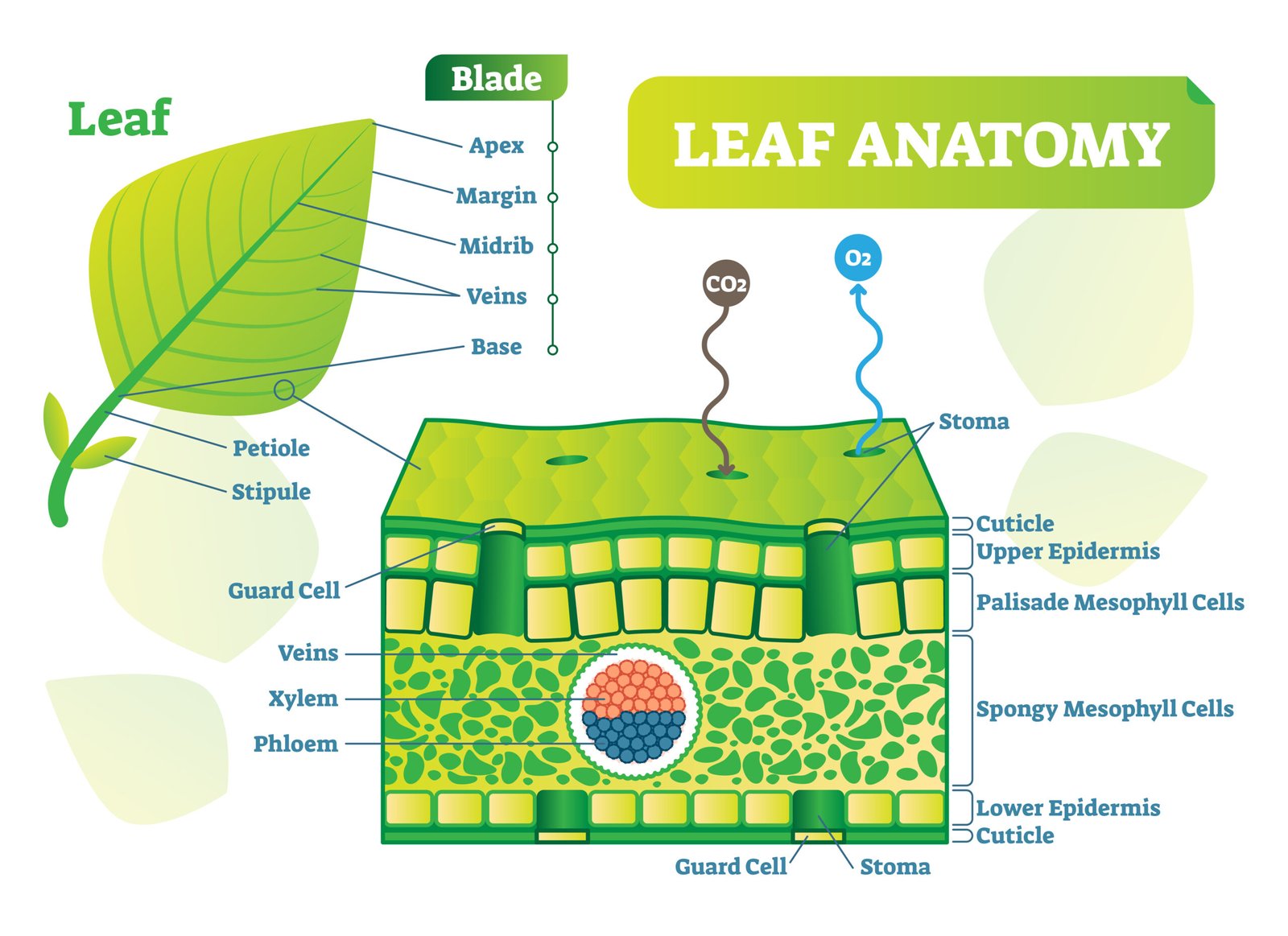
🔵 Question 7. Describe the internal structure of a dorsiventral leaf with the help of labelled diagrams.
🟢 Answer:
🌿 Dorsiventral leaf (typical dicot leaf, e.g. mustard):
Upper epidermis: single layer, cuticle, no chloroplast.
Mesophyll:
➡️ Palisade parenchyma — upper side, elongated cells, rich in chloroplasts.
➡️ Spongy parenchyma — lower side, air spaces.
Vascular bundles: Conjoint, collateral; xylem towards upper, phloem towards lower.
Lower epidermis: with stomata for gaseous exchange.
✏️ Labelled diagram description: Upper epidermis → palisade → spongy → lower epidermis with stomata → vascular bundle in centre.
✔️ Function: Efficient photosynthesis and gaseous exchange.
————————————————————————————————————————————————————————————————————————————
OTHER IMPORTANT QUESTIONS FOR EXAMS
(CBSE MODEL QUESTIONS PAPER)
ESPECIALLY MADE FROM THIS LESSON ONLY
🔴 Question 1:
The study of internal structure of plants is called:
🔴1️⃣ Morphology
🟢2️⃣ Anatomy
🟡3️⃣ Cytology
🔵4️⃣ Histology
🟢 Answer: 2️⃣ Anatomy
🔴 Question 2:
The permanent tissues are derived from:
🔴1️⃣ Apical meristem
🟢2️⃣ Lateral meristem
🟡3️⃣ Meristematic tissues
🔵4️⃣ Intercalary meristem
🟢 Answer: 3️⃣ Meristematic tissues
🔴 Question 3:
Apical meristem is responsible for:
🔴1️⃣ Secondary growth
🟢2️⃣ Primary growth
🟡3️⃣ Increase in girth
🔵4️⃣ Healing of wounds
🟢 Answer: 2️⃣ Primary growth
🔴 Question 4:
The thickening of cell wall due to deposition of lignin is found in:
🔴1️⃣ Collenchyma
🟢2️⃣ Parenchyma
🟡3️⃣ Sclerenchyma
🔵4️⃣ Xylem parenchyma
🟢 Answer: 3️⃣ Sclerenchyma
🔴 Question 5:
Xylem and phloem together constitute:
🔴1️⃣ Simple tissue
🟢2️⃣ Complex tissue
🟡3️⃣ Meristematic tissue
🔵4️⃣ Protective tissue
🟢 Answer: 2️⃣ Complex tissue
🔴 Question 6:
Which element of xylem is living?
🔴1️⃣ Vessels
🟢2️⃣ Tracheids
🟡3️⃣ Xylem fibres
🔵4️⃣ Xylem parenchyma
🟢 Answer: 4️⃣ Xylem parenchyma
🔴 Question 7:
Phloem in gymnosperms lacks:
🔴1️⃣ Sieve tubes
🟢2️⃣ Companion cells
🟡3️⃣ Phloem parenchyma
🔵4️⃣ Phloem fibres
🟢 Answer: 2️⃣ Companion cells
🔴 Question 8:
The cork cells are impervious to water due to the presence of:
🔴1️⃣ Cellulose
🟢2️⃣ Lignin
🟡3️⃣ Suberin
🔵4️⃣ Cutin
🟢 Answer: 3️⃣ Suberin
🔴 Question 9:
The secondary growth in dicot stem is due to:
🔴1️⃣ Apical meristem
🟢2️⃣ Intercalary meristem
🟡3️⃣ Vascular cambium and cork cambium
🔵4️⃣ Pith cambium
🟢 Answer: 3️⃣ Vascular cambium and cork cambium
🔴 Question 10:
The radial arrangement of vascular bundles is found in:
🔴1️⃣ Dicot stem
🟢2️⃣ Monocot stem
🟡3️⃣ Dicot root
🔵4️⃣ Monocot leaf
🟢 Answer: 3️⃣ Dicot root
🔴 Question 11:
Define simple permanent tissues and name their types.
🟢 Answer:
Definition: Simple tissues are made of similar cells performing same function.
Types:
1️⃣ Parenchyma: Living, thin-walled, stores food.
2️⃣ Collenchyma: Living, thick corners, mechanical support.
3️⃣ Sclerenchyma: Dead, lignified, gives rigidity.
🔴 Question 12:
What are complex permanent tissues? Give two examples.
🟢 Answer:
Definition: Composed of different types of cells working together.
Examples:
1️⃣ Xylem — transports water 💧.
2️⃣ Phloem — transports food 🍞.
🔴 Question 13:
What are meristematic tissues? Describe their types based on position.
🟢 Answer:
Definition: Tissues with actively dividing cells, responsible for plant growth.
Types (based on position):
1️⃣ Apical meristem:
• Found at tips of root and shoot.
• Responsible for primary growth (length).
2️⃣ Intercalary meristem:
• Found at base of nodes or leaves.
• Causes regrowth in grasses 🌾.
3️⃣ Lateral meristem:
• Present along sides of stem and root.
• Responsible for secondary growth (girth).
🔴 Question 14:
Describe the simple permanent tissues and their functions.
🟢 Answer:
1️⃣ Parenchyma:
• Living cells; thin walls; large vacuoles.
• Functions: storage, photosynthesis, healing.
2️⃣ Collenchyma:
• Living, thickened corners with cellulose.
• Provides mechanical support & flexibility.
3️⃣ Sclerenchyma:
• Dead, lignified walls.
• Provides hardness & rigidity.
🧠 Together, they form basic permanent tissues performing simple functions.
🔴 Question 15:
Write the main components and functions of xylem.
🟢 Answer:
Components:
1️⃣ Tracheids — water conduction & support.
2️⃣ Vessels — main conducting elements 💧.
3️⃣ Xylem fibres — mechanical strength.
4️⃣ Xylem parenchyma — storage.
Function:
✔️ Transports water & minerals upward.
✔️ Provides mechanical support.
🔴 Question 16:
Write the main components and functions of phloem.
🟢 Answer:
Components:
1️⃣ Sieve tube elements — conduction of food.
2️⃣ Companion cells — control activities of sieve tubes.
3️⃣ Phloem parenchyma — stores food.
4️⃣ Phloem fibres — mechanical support.
Function:
✔️ Transports organic food (sucrose) from leaves 🍃 to other parts.
🔴 Question 17:
Differentiate between open and closed vascular bundles.
🟢 Answer:
Feature Open Closed
Cambium Present Absent
Secondary growth Occurs No secondary growth
Example Dicot stem 🌻 Monocot stem 🌾
💡 Open bundles show secondary growth due to cambium.
🔴 Question 18:
Describe the anatomy of dicot stem 🌻.
🟢 Answer:
Epidermis: Outermost layer with cuticle.
Cortex: Collenchyma + parenchyma.
Endodermis: Starch sheath.
Pericycle: Few layers beneath endodermis.
Vascular bundles: Arranged in ring, open & conjoint.
Pith: Large central parenchyma.
Feature: Shows secondary growth.
🔴 Question 19:
Describe the anatomy of monocot stem 🌾.
🟢 Answer:
Epidermis: Outer layer with thick cuticle.
Ground tissue: Uniform, no cortex-pith distinction.
Vascular bundles: Scattered, closed & conjoint.
Bundle sheath: Present.
Feature: No secondary growth.
🔴 Question 20:
Describe the anatomy of dicot root.
🟢 Answer:
Epiblema: Outermost with root hairs.
Cortex: Parenchymatous.
Endodermis: Casparian strips.
Pericycle: Gives rise to lateral roots.
Vascular bundles: Radial, xylem exarch.
Pith: Small or absent.
Feature: Shows secondary growth.
🔴 Question 21:
Describe the anatomy of monocot root 🌽.
🟢 Answer:
Epiblema: Outer layer with root hairs.
Cortex: Many parenchyma layers.
Endodermis: Casparian strips.
Pericycle: Gives lateral roots.
Vascular bundles: Radial, polyarch, xylem exarch.
Pith: Large & well-developed.
Feature: No secondary growth.
🔴 Question 22:
Differentiate between monocot and dicot root.
🟢 Answer:
Feature Dicot Root Monocot Root
Vascular bundles 4–6 (few) Many (polyarch)
Pith Small/absent Large
Secondary growth Present Absent
Example Bean 🌿 Maize 🌾
🔴 Question 23:
Describe the secondary growth in dicot stem 🌻 with stages.
🟢 Answer:
Definition: Formation of secondary tissues by lateral meristems (cambium) increases the girth of stem.
Process:
1️⃣ Formation of cambium ring:
• In vascular bundles, intrafascicular cambium is joined by interfascicular cambium to form a continuous ring.
2️⃣ Activity of cambium:
• Produces secondary xylem inward and secondary phloem outward.
3️⃣ Formation of annual rings:
• Alternating spring and autumn wood form growth rings 🌳.
4️⃣ Cork cambium (phellogen):
• Arises from cortex → produces phellem (cork) outside and phelloderm inside.
5️⃣ Periderm formation:
• Phellem + phellogen + phelloderm = Periderm.
Result:
✅ Increase in girth.
✅ Protection through cork layer.
🔴 Question 24:
Describe secondary growth in dicot root 🌱.
🟢 Answer:
Initiation: Secondary growth starts from cambium formed by pericycle and conjunctive tissue.
Steps:
1️⃣ Cambium formation: Conjunctive tissue cells become cambial strips.
2️⃣ Cambium activity: Forms secondary xylem inward, secondary phloem outward.
3️⃣ Cork cambium: Formed from pericycle; produces cork externally.
4️⃣ Result: Root becomes thick due to secondary tissues.
Feature:
✔️ Annual rings absent.
✔️ Function: Strength and conduction.
🔴 Question 25:
Explain the structure and functions of epidermis in plants.
🟢 Answer:
Structure:
• Outermost layer of cells covering all plant parts.
• Usually single-layered, with cuticle on aerial parts.
• May have trichomes and stomata.
Functions:
1️⃣ Protection from mechanical injury and infection.
2️⃣ Reduces water loss by cuticle.
3️⃣ Exchange of gases through stomata.
4️⃣ Absorption in roots (root hairs 💧).
🔴 Question 26:
Describe the structure of stomata and lenticels and their roles.
🟢 Answer:
Stomata:
1️⃣ Pores on epidermis surrounded by guard cells.
2️⃣ Present in leaves 🍃; control transpiration and gas exchange.
3️⃣ Open/close due to turgor pressure.
Lenticels:
1️⃣ Openings in cork layer.
2️⃣ Made of loosely arranged parenchyma.
3️⃣ Help in gaseous exchange during secondary growth.
💡 Both maintain internal gaseous balance.
🔴 Question 27:
Write differences between primary xylem and secondary xylem.
🟢 Answer:
Feature Primary Xylem Secondary Xylem
Origin From procambium From vascular cambium
Function Conduction in young plant Conduction + support
Position Near centre Between primary xylem and phloem
Growth rings Absent Present
Secondary xylem forms annual rings 🌳 in dicot stem.
🔴 Question 28:
Explain the structure and function of periderm.
🟢 Answer:
Definition: Protective tissue formed during secondary growth.
Components:
1️⃣ Phellogen (cork cambium): Meristematic, produces other two layers.
2️⃣ Phellem (cork): Outer dead layer with suberin, water-proof.
3️⃣ Phelloderm: Inner living parenchyma.
Function:
✔️ Replaces epidermis.
✔️ Prevents water loss.
✔️ Provides protection.
🔴 Question 29:
Differentiate between heartwood and sapwood.
🟢 Answer:
Feature Heartwood Sapwood
Color Dark Light
Function Non-conducting Conducting
Position Central Peripheral
Role Support Conduction of water 💧
Heartwood: Durable and resistant to decay.
Sapwood: Functional in water transport.
🔴 Question 30:
Write short notes on growth rings and bark.
🟢 Answer:
Growth rings:
• Alternate layers of spring wood (lighter) and autumn wood (darker) formed due to seasonal activity of cambium.
• Each ring = 1 year’s growth.
• Used to determine age of plant 🌳.
Bark:
• All tissues external to vascular cambium.
• Early bark: Formed early in season.
• Late bark: Formed later; thicker and protective.
————————————————————————————————————————————————————————————————————————————